-
Pawar HospitalPawar Hospital (+91) 9175898623
-
Spire HospitalSpire Hospital (+91) 9175983868
-
Book AppointmentBook Appointment
Dr. Gaurav Pawar is a well established Hip and Knee joint replacement and sports injury (Arthroscopy) surgeon. He has eight years of experience in Trauma & Orthropaedics and has gained considerable knowledge and espertise in this field during this period. He has also worked in Trauma and Orthopaedics in the NHS system in UK for 3 years at reputed hospitals like Royal Free Hospital,London, Chase Farm Hospital, London and Tameside Hospital, Greater Manchester. He has performed numerous joint replacement and arthroscopy surgeries during this period and is well versed with the latest surgical techniques. He has also presented and published many papers at national and international stages.
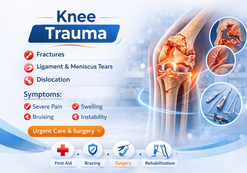
Knee Trauma
Knee Trauma
Femur & Tibial Fracture Fixation
Femur Fracture Fixation
Distal Femur Screws
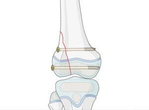
Distal femur screws are orthopedic implants used in the treatment of fractures and other conditions affecting the distal (lower) part of the femur, which is the thigh bone. The distal femur is the area near the knee joint. These screws are designed to provide stability and support to the fractured bone, allowing for proper healing.
Fractures of the distal femur can occur due to various reasons, such as trauma, accidents, or osteoporosis. When a fracture occurs, especially in a weight-bearing bone like the femur, it’s important to immobilize the fractured bone and promote proper alignment for healing. Distal femur screws play a crucial role in this process.
The screws used for this purpose are typically made of materials like stainless steel or titanium, which are biocompatible and can remain in the body without causing adverse reactions. The screws come in various sizes and shapes to accommodate different fracture patterns and patient anatomy.
Distal Femur Nail
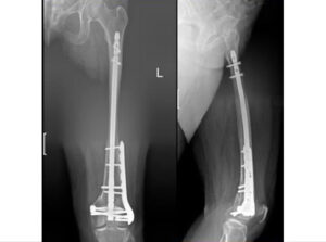
A distal femur nail, also known as a distal femoral nail or distal femur intramedullary nail, is an orthopedic implant used to treat fractures of the distal (lower) part of the femur, near the knee joint. This type of implant is designed to provide stabilization and support to the fractured bone, allowing for proper healing and alignment.
Here’s how the distal femur nail works and some key points about its use:
Surgical Procedure: The surgical procedure involves making an incision near the knee joint and inserting the intramedullary nail into the marrow cavity of the femur. The nail is typically inserted from the top of the femur (proximal) and guided down to the fractured area (distal) using fluoroscopy (real-time X-ray) guidance.
Nail Design: The nail is usually made of materials like stainless steel or titanium, which are strong and biocompatible. The nail has a tapered or cylindrical design that allows it to fit within the intramedullary canal of the bone. It may have holes along its length for screws or locking bolts to secure it in place and stabilize the fracture.
Distal Femur Plate
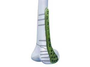
A distal femur plate is an orthopedic implant used to treat fractures of the distal (lower) part of the femur, which is the thigh bone. This type of implant is designed to provide stability and support to fractured bone fragments, allowing for proper healing and alignment. Distal femur plates are commonly used in cases where the fracture is too complex or displaced to be treated effectively with other methods, such as casts or intramedullary nails.
Here are some key points about distal femur plates:
Surgical Procedure: The surgical procedure involves making an incision near the knee joint and placing the plate on the surface of the bone. The plate is secured to the bone using screws, which are inserted through holes in the plate and into the bone fragments. The plate provides a stable fixation and holds the bone fragments in proper alignment during the healing process.
Plate Design: Distal femur plates come in various shapes and sizes to accommodate different fracture patterns and patient anatomy. They are typically made of materials like stainless steel or titanium, which are strong, biocompatible, and can remain in the body without causing adverse reactions.
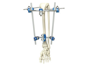
Distal Femur External Fixator & Knee Replacement
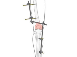
A distal femur external fixator is a medical device used to stabilize and treat fractures of the distal (lower) part of the femur, which is the thigh bone. Unlike internal fixation methods like plates, screws, and nails, an external fixator is applied outside the body and provides stability to the fractured bone fragments through the use of pins or wires inserted into the bone.
Here are some key points about distal femur external fixators:
Purpose: External fixators are often used when internal fixation methods are not suitable due to the severity of the fracture, soft tissue damage, or infection. They can also be used for temporary stabilization before more definitive surgery is performed.
Components: A typical external fixator consists of metal pins or wires that are inserted into the bone fragments above and below the fracture site. These pins are connected by a rigid frame that holds the bone in proper alignment. The frame is positioned outside the body, and it can be adjusted to provide the desired stability.
Tibial Fracture Fixation
Tibial fracture fixation is a vital orthopedic procedure designed to address fractures in the tibia, or shin bone. This surgical intervention aims to stabilize and align the broken bone segments using specialized implants such as screws, intramedullary nails, or plates. The primary objective is to promote proper bone healing, restore structural integrity, and facilitate a successful recovery.
During the procedure, orthopedic surgeons strategically place implants within or alongside the fractured bone fragments. Screws can be employed to hold the bone fragments in place, while intramedullary nails are inserted into the bone’s marrow canal to provide internal stabilization. Plates are utilized to offer additional support and alignment along the bone’s surface.
Proximal Tibia Screws
Proximal Tibia Nail
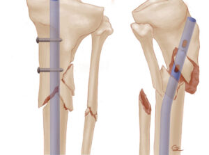
A proximal tibia nail is a sophisticated orthopedic implant designed to address fractures and injuries occurring in the upper portion of the tibia, the shin bone, near the knee joint. This innovative implant serves as a crucial element in modern orthopedic practice, offering a comprehensive solution for stabilizing fractures and promoting optimal healing outcomes.
The procedure involving a proximal tibia nail entails a surgical intervention performed under sterile conditions. An incision is made near the knee joint to access the fractured area. The nail, typically composed of biocompatible materials like stainless steel or titanium, is then inserted into the marrow cavity of the tibia. The insertion process is guided by fluoroscopy, a real-time X-ray imaging technique that aids in precise nail placement. The nail’s design, often tapered or cylindrical, enables it to fit within the intramedullary canal of the bone, providing internal support while minimizing disruption to the surrounding soft tissues.
Proximal tibia nails possess locking mechanisms at both ends, allowing for the insertion of screws or locking bolts that engage with the nail. This locking mechanism provides enhanced stability, preventing rotational movement of the bone fragments and enabling controlled healing. The procedure strives to restore the bone’s anatomical alignment and promote fracture union through controlled compression and load-sharing properties.
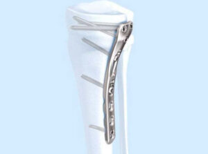
Proximal Tibia Plate
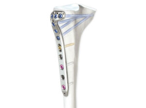
A proximal tibia plate is a specialized orthopedic implant designed to address fractures and injuries in the upper part of the tibia, the shin bone, near the knee joint. This implant plays a crucial role in stabilizing fractured bone fragments and facilitating proper healing.
During the surgical procedure, a proximal tibia plate is affixed to the surface of the bone through an incision near the knee joint. The plate is secured using screws that pass through holes in the plate and anchor into the bone. This fixation provides stability to the fractured area, allowing for optimal alignment and healing.
Proximal tibia plates are meticulously designed to accommodate various fracture patterns and patient anatomies. Constructed from biocompatible materials like stainless steel or titanium, these plates ensure compatibility with the body while maintaining structural integrity.
Proximal Tibia External Fixator & Knee Replacement
Sports Injury
Physiotherapy & Rehabilitation